Magnetic resonance imaging (MRI)
About the facility
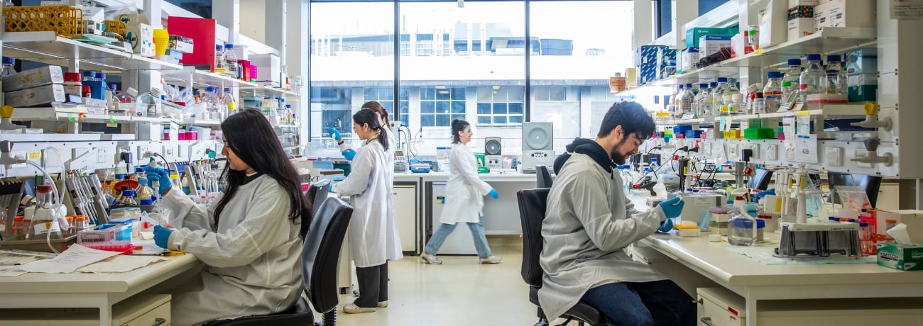
The research MRI facilities at The Florey facilitate cross-disciplinary collaboration between clinicians and scientists across Melbourne. Our imaging platform is equipped to support the acquisition of cutting-edge human neuroimaging in clinical populations from across the lifespan, including high-resolution neuroanatomical, functional and diffusion imaging.
Located on the ground floor of the Melbourne Brain Center at the Austin Hospital Heidelberg, the MRI facility is co-located with several Florey and University of Melbourne research laboratories and specialised Austin Hospital clinics.
The MRI facility is available for use by the imaging research community Australia-wide to carry out advanced imaging studies.
3T MRI
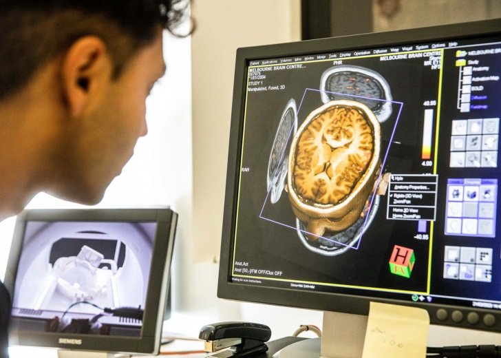
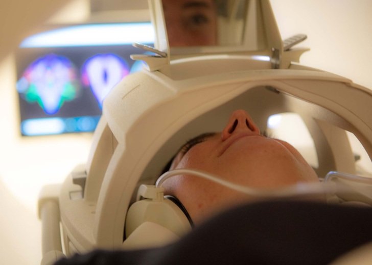
The Florey’s MRI facility is unique in Australia, housing state-of-the-art 3-tesla MRI scanners dedicated to research.
Our instrumentation includes a Siemens 3T Prisma Fit MRI and a Siemens 3T wide-bore Vida MRI. These MRI scanners are complemented by a range of peripheral equipment including high density EEG, transcranial magnetic stimulation, in-scanner eye-tracking, prospective motion correction and phantoms designed for precision quantitative imaging.
The facility also houses a mock MRI scanner that is particularly useful for preparing children or those nervous about an MRI for their scan.
MRI-guided focused ultrasound (MRgFUS)
MRI-guided focused ultrasound (MRgFUS) is an incisionless medical procedure used for treating neurological disorders.
Using the MRgFUS technology, Florey researchers aim to develop personalised treatment strategies for people with brain network disorders.
Learn moreMRI imaging partners
The Florey’s Heidelberg MRI facility is a node of the National Imaging Facility.
The NIF network facilitates access to imaging equipment, expertise, and tools to solve critical problems related to human health and disease.
Imaging software
Florey imaging researchers have developed a range of image analysis software, free to download. This includes iBrain™ and the The Integrated Brain Analysis Toolbox for SPM (iBT), which can work independently or together to perform neuroimage processing, analysis and visualisation.
Contact
Chris Kokkinos
Lead Research Radiographer
E [email protected]